



Grade 1 Spondylolisthesis: Causes, Treatments, Recovery Forecasts
Grade 1 spondylolisthesis, defined by a 0% to 25% forward vertebral slippage, is the mildest form of spondylolisthesis. Also known as low-grade spondylolisthesis, it results from spinal instability from age-related spinal degeneration or spinal stress fractures (called spondylolysis) from overuse. Since it’s the least severe form of the condition, grade 1 spondylolisthesis is usually resolved with conservative methods, although it still requires prompt diagnosis and treatment to prevent worsening back pain, instability, and nerve compression.
In this comprehensive guide, we’ll explore spondylolisthesis grading systems with specific causes, effective treatment options, and a clear recovery forecast to lead you toward a mobile, pain-free life.

What Does Grade 1 Spondylolisthesis Mean?
Grade 1 spondylolisthesis means that the affected vertebra has slipped between 1% and 25%, according to the Meyerding classification system. It’s the mildest and most common form of vertebral slippage.
Meyerding Classification for Grade 1 Spondylolisthesis
The Meyerding classification system of spondylolisthesis categorizes vertebral slippage severity into five grades using a lateral spine X-ray image.
To use the Meyerding classification, the provider draws two vertical lines along the posterior cortex superior and inferior vertebrae in the lateral spine X-ray. They measure the distance between them (A) and the length of the interior vertebral body (B), then calculate A/B to arrive at the percentage that corresponds to a grade.
In X-ray images displaying flexion or extension views of the spine, a vertebral slippage of greater than four millimeters signals instability.
| Meyerding Classification for Spondylolisthesis | |
| Grade | Degree of Slippage |
| Grade I | 0% to 25% |
| Grade II | 25% to 50% |
| Grade III | 50% to 75% |
| Grade IV | 75% to 100% |
| Grade V (Spondyloptosis) | Over 100% |
What is Spondylolisthesis? Understanding Types and Etiology
Spondylolisthesis occurs when one of the spinal bones, called a vertebra, slips forward out of normal alignment onto the bone beneath it. It can be separated into five types using the Wiltse classification system: dysplastic (congenital), isthmic, degenerative, traumatic, and pathologic.
| Wiltse Classification for Spondylolisthesis | |
| Type | Etiology (Cause) |
| Type I: Dysplastic/Congenital | Genetic abnormality in the spinal facet joints |
| Type II: Isthmic | Defect in the pars interarticularisSubtype A (lytic): Caused by a pars stress fracture (spondylolysis)Subtype B (elongated pars): Caused by multiple healed injuries, causing the pars to elongateSubtype C (acute pars fracture): Caused by a single traumatic event to the pars |
| Type III: Degenerative | Progressive degenerative spinal changes |
| Type IV: Traumatic | A sudden, acute injury to the spine |
| Type V: Pathologic | Underlying bone disease, like an infection or Paget’s disease |
Isthmic (type II) and degenerative (type III) are the most common types of spondylolisthesis. Some sources also include a sixth type of spondylolisthesis in the Wiltse classification system: iatrogenic, or post-surgical. This refers to spondylolisthesis that occurs after a medical treatment.
H4: Spondylolisthesis vs. Spondylolysis
Spondylolisthesis refers to a forward slippage of a vertebra, while spondylolysis is a stress fracture in the pars interarticularis, the bone segment that connects two facet joints between vertebrae. Spondylolysis can cause isthmic spondylolisthesis.
The Difference Between Isthmic and Degenerative Slips
- Cause: Degenerative spondylolisthesis is caused by ongoing spinal wear and tear; isthmic spondylolisthesis is caused by a defect in the pars interarticularis (usually a stress fracture, called spondylolysis).
- Age: Degenerative spondylolisthesis usually occurs after age 60, while isthmic spondylolisthesis often develops in kids and teens, starting from age six, according to a Global Spine Journal study.
- Location: Degenerative spondylolisthesis most commonly affects the L4-L5 spinal level, while isthmic spondylolisthesis often affects the L5-S1 level, according to Spondylolisthesis, published in StatPearls.
Detailed Symptoms and Clinical Presentation
Common symptoms:
- Lower back pain that tends to worsen with standing and walking and improve with sitting or leaning forward
- Neurogenic claudication, a collection of symptoms caused by nerve compression in the lumbar spine that may affect the lower back and one or both legs, buttocks, and hips. It tends to worsen when walking or standing upright and improve with sitting or leaning forward. Symptoms include:
- Pain
- Tingling
- Cramping
- Numbness
- Leg weakness or heaviness with walking
- Muscle spasms and tightness, particularly in the hamstrings
- Sciatica, pain along the path of the sciatic nerve, which travels from the lower back through the buttocks and down the backs of both legs.
- Unlike mechanical pain, nerve pain from spondylolisthesis often radiates from the lower back to the legs. Nerve pain tends to be described as sharp, shooting, or burning.
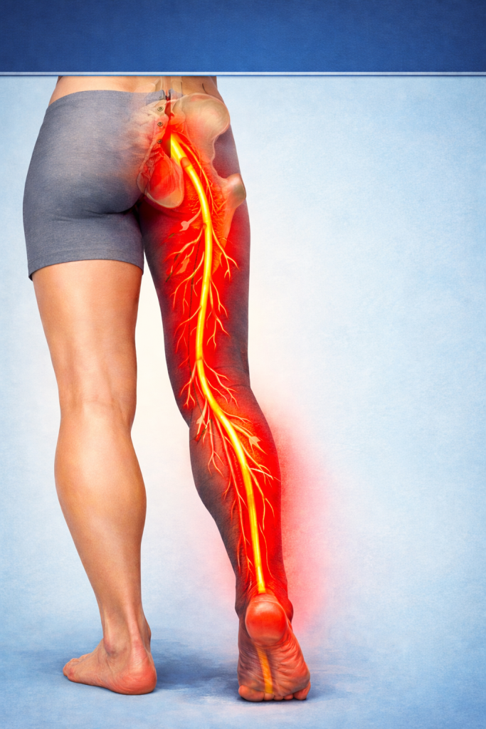
While the above symptoms can occur if the vertebral slippage compresses nearby nerves, many cases of grade 1 spondylolisthesis are asymptomatic, meaning they don’t produce any noticeable symptoms.
Red Flag Symptoms: When to Get Immediate Medical Attention
While rare with grade 1 spondylolisthesis, more advanced forms of spondylolisthesis can lead to severe nerve compression and potentially cauda equina syndrome, a medical emergency resulting from compression of a small bundle of nerves at the base of the spine. If you experience these cauda equina syndrome symptoms, head to the emergency room for immediate care:
- Saddle anesthesia (numbness in the inner thighs, buttocks, and perineum
- Bladder or bowel dysfunction
- Sexual dysfunction
- Leg weakness or numbness
Is Grade 1 Spondylolisthesis Serious?
Grade 1 spondylolisthesis is considered the least serious form of this spinal condition. Additionally, grade 1 and grade 2 are the most frequently diagnosed forms of spondylolisthesis. However, if the condition isn’t identified early on, or if the patient doesn’t follow an effective treatment plan, spondylolisthesis can progress.
What Are The Causes and Risk Factors for Grade 1 Spondylolisthesis?
Risk factors for grade 1 spondylolisthesis include spinal wear and tear, age-related spinal changes, and genetic abnormalities, like thin bone tissue and neural arch defects.
Age-Related Spinal Wear and Tear (Degenerative)
Age-related spinal wear and tear can cause degenerative spondylolisthesis, the most common form of vertebral slippage in older adults. Degenerative spondylolisthesis most often occurs at the L4-L5 spinal level.
Various factors can subject the spine to wear and tear throughout a lifetime, including:
- High-impact activities, like running and contact sports
- Repetitive spinal bending, twisting, or lifting
- Poor posture
- Being overweight or obese
Aspects of the natural aging process also contribute to age-related spinal wear and tear. The spine inevitably changes with age: the intervertebral discs that absorb impact to protect the spine become thinner and drier. Bone mass reduces with age, leading to thinner spinal structures that are more susceptible to injury.
All of these factors can contribute to the development of degenerative grade 1 spondylolisthesis, which typically affects patients over the age of 60.
Overextending The Spine (Isthmic)
Certain sports involve repeatedly extending the spine and involve a high risk of spinal injury, including gymnastics, volleyball, diving, and football. Young athletes who participate in these sports are more likely to develop grade 1 isthmic spondylolisthesis from a pars stress fracture. While this can occur at any spinal level, it’s most common at L5-S1.
Genetic Factors and Structural Anomalies (Congenital)
Some individuals are more likely to develop spondylolisthesis due to genetic factors present at birth, such as:
- Thin bone tissue: Some people naturally have thinner bone tissue in the vertebrae from abnormal bone formation, leading to a higher risk of spinal injury.
- Neural arch defect: This spondylolisthesis type usually stems from a neural arch defect at L5-S1 (the neural arch is the bony arch of a vertebra surrounding the spinal cord).
- Spina bifida occulta: 95% of congenital spondylolisthesis cases are linked to spina bifida occulta, a birth defect with which the back of the spine doesn’t fully enclose the spinal cord, according to MedScape.
How is Grade 1 Spondylolisthesis Diagnosed?
Grade 1 spondylolisthesis is typically diagnosed through a combination of physical exams, neurological exams, and imaging tests.
Exams for Grade 1 Spondylolisthesis
The examination process for grade 1 spondylolisthesis typically involves physical and neurological evaluations.
Physical examinations: Your doctor may gently move your legs in different positions to see if any specific movements cause pain or neurological symptoms. They may also gently press on the spine to check for pain or tenderness and have you walk around to evaluate your gait.
- Goal: These manual techniques can help your provider identify the location of the vertebral slip and which nerves are affected. They can also offer insights into how advanced the condition is based on its effect on your posture, movement, balance, and gait.
Neurological examinations: Your doctor will check your nerve reflexes to identify which muscles are affected by spinal nerve compression. They may use a small hammer to identify numb nerve signals or place stimuli (like heat or cold) on the skin.
- Goal: Identify the affected spinal segment.
Imaging Tests to Diagnose Grade 1 Spondylolisthesis
Imaging tests allow your doctor to visualize the spine after evaluating your symptoms. They can check for a slipped vertebra, determine the grade of the slippage, and check for nerve damage. Options include X-rays, CT scans, MRI scans, and myelograms.
X-rays are often the first imaging tests that doctors order to diagnose spondylolisthesis. They use electromagnetic waves to create pictures of the inside of the body. X-rays are primarily used to examine bones and joints – in the case of spondylolisthesis, they can display the slipped vertebra.
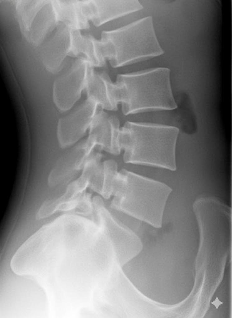
Diagnosing Instability: The Role of Dynamic X-Rays
Providers often use dynamic imaging, which captures multiple photos to illustrate the body in motion, to evaluate whether spondylolisthesis is dynamic (unstable) or static (stable). If the forward or backward movement of the vertebra (called sagittal translation) is greater than 4 mm, or if it exhibits an angular change of more than 10° on lateral-flexion radiographs, the instability is considered significant.
Other Imaging Tests
- CT scans: Your doctor may implement a CT scan if an X-ray shows that a vertebra has slipped forward, but they need a more detailed view of the spine. CT scans use computerized X-rays that rapidly rotate around the patient, creating detailed cross-sectional images of the body at different angles. These images can provide additional information about a pars interarticularis fracture, a possible cause of spondylolisthesis.
- MRI scans use a combination of strong magnets and radiofrequency energy to create high-resolution images of the body. This advanced imaging test is especially helpful for viewing soft tissue, including the spinal cord, nerves, muscles, and ligaments, in great detail. Your physician may order an MRI while diagnosing spondylolisthesis to identify possible spinal nerve impingement.
- Myelograms involve injecting a contrast dye into the spinal column before taking an X-ray or CT scan. The dye allows your healthcare provider to view the spinal cord in greater detail than with an X-ray or CT scan alone. Spinal myelograms can identify compression of the spinal cord or nerves, which occurs in some spondylolisthesis cases.
Imaging tests are a valuable tool in determining the grade of vertebral slippage for spondylolisthesis diagnoses. Often, grade-1 spondylolisthesis is mild enough to be classified with an X-ray alone, without the need for an MRI or more extensive testing. However, your physician will make personalized recommendations based on your spondylolisthesis symptoms and general health.
Comprehensive Non-Surgical Treatment Protocol
Non-surgical treatment protocols for grade 1 spondylolisthesis typically include rest, activity modifications, physical therapy, and anti-inflammatory medications. If required, the patient may also undergo steroid injections or spinal bracing, which help prevent spinal instability.
Rest and Activity Modifications
- Avoid high-impact activities like weightlifting, gymnastics, contact sports, and yoga poses involving extreme spinal bending or twisting.
- Strengthen your core muscles through targeted, low-impact exercises.
- Improve workplace ergonomics by using a chair with robust lumbar support and avoiding prolonged periods of sitting.
- Prioritize good posture to keep the spine in neutral alignment.
Medication and Injections
- Over-the-counter NSAIDs (non-steroidal anti-inflammatory drugs) are often used for grade 1 spondylolisthesis. Prescription NSAIDs may be required in more severe cases.
- Muscle relaxants may help with muscle spasms caused by spondylolisthesis.
- Steroid injections may be prescribed in more severe cases to manage pain and inflammation, although their effects are temporary.
Physical Therapy (PT) and Core Stabilization
Physical therapy goals for grade 1 spondylolisthesis include:
- Reducing pain levels and nerve symptoms
- Improving spinal stability
- Building core strength
- Improving and retaining mobility
- Correcting body mechanics to prevent worsening instability and slippage
To achieve this, your PT may provide:
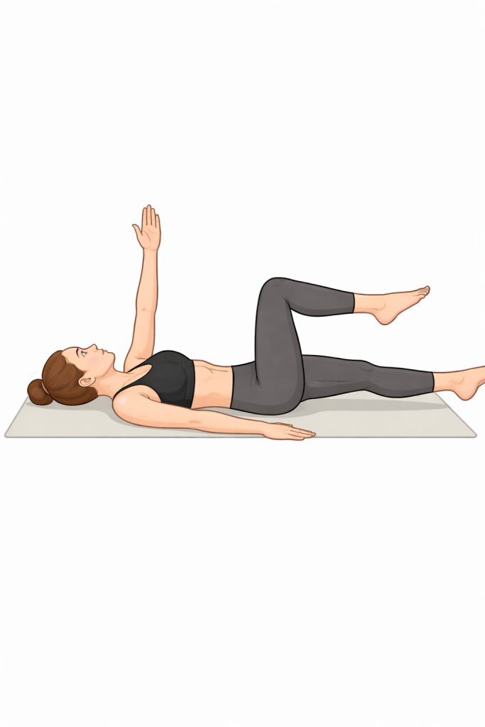
- Targeted core strengthening exercises, including:
- Pelvic tilts
- Bird-dogs
- Modified planks
- Bridges
- Curl-ups
- Dead bug
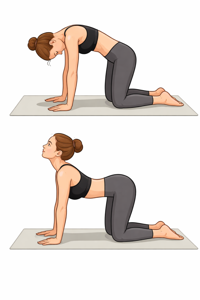
- Targeted stretches for mobility, including:
- Knee-to-chest
- Child’s pose
- Seated hamstring stretch
- Cat-cow
- Posture assessments and movement pattern guidance
- Complementary treatments to naturally reduce pain, such as:
- Heat/cold therapy
- Massage therapy
- Acupuncture or dry needling
- Biofeedback
- Cupping
- Kinesio taping
Specific Exercises to Avoid and Safe Alternatives
Safe, low-impact forms of exercise promote spinal mobility and muscle strength. Some of the best forms of exercise for grade 1 spondylolisthesis include:
- Walking
- Swimming and water aerobics
- Using an elliptical machine
- Certain strengthening exercises, such as side planks and pelvic tilts
- Gentle stretching
- Non-intense cycling
Certain forms of exercise can lead to spondylolisthesis progression and worsened symptoms. These include:
- Weightlifting
- Some sports, including wrestling, gymnastics, football, and other contact sports
- Yoga poses that involve back extension
- Diving
- Back extensions
- Sit-ups and crunches
- Any activity that involves significantly twisting or rotating the spine
Ultimately, you should talk to your doctor about which forms of exercise to participate in with grade 1 spondylolisthesis. As your back heals, your doctor may give you the go-ahead to partake in a wider range of physical activities.
H4: Can I Run With Grade 1 Spondylolisthesis?
If you have grade 1 spondylolisthesis and are experiencing symptoms, it’s best to avoid running until your doctor clears you to do so. Running is a high-impact activity that places extra stress on the spine and may cause worsened back pain and neurological symptoms.
Bracing
Back bracing is typically used for acute pain and instability in kids and teens with isthmic grade 1 spondylolisthesis from a stress fracture. Spondylolisthesis braces and corsets compress the torso to stabilize and alleviate pressure from the spine. By preventing potentially damaging movements, the brace can allow the spine to heal.
Keep in mind that bracing provides short-term support – it’s not a long-term cure for spondylolisthesis.
Surgical Options: When Conservative Care Fails
When conservative methods fail to alleviate spondylolisthesis symptoms, your provider may present surgical options, including spinal decompression and spinal fusion.
Does Grade 1 Spondylolisthesis Need Surgery?
Grade 1 spondylolisthesis generally doesn’t require surgery. It’s the mildest form of the condition, so its symptoms can typically be alleviated with non-invasive methods. But spine specialists may recommend surgery for grade 1 spondylolisthesis if:
- Non-surgical treatment methods fail to provide relief after an extended period (typically six months or longer)
- Significant neurological deficits are present
- Surgery is required to prevent the condition’s progression
- The altered alignment of the spine severely impacts the patient’s function and posture
Note that grade 1 spondylolisthesis can’t be cured without surgery – once the vertebra has slipped out of its normal position, surgery is the only way to restore the alignment of the spine.
The Goals of Surgery
| Spondylolisthesis Surgery Treatment Goals | ||
| Decompression | Spinal Fusion | |
| Overview | Remove tissue that’s causing nerve impingement, usually lamina or disc material | Place bone graft material between affected vertebrae to permanently fuse them |
| Goal | Alleviate nerve compression symptoms | Stabilize the spine |
| Approaches | LaminectomyDiscectomyMicrodiscectomyForaminotomyMinimally invasive decompression | Posterior lumbar interbody fusion (PLIF)Anterior lumbar interbody fusion (ALIF)Lateral lumbar interbody fusion (LLIF/XLIF) |
Traditional Surgical Approaches: Decompression and Spinal Fusion
Spinal decompression is traditionally performed with spinal fusion to resolve instability from spondylolisthesis. According to the Journal of Orthopaedics, fusion has been around for over a century, making it a time-tested procedure for spinal conditions. But like any surgical procedure, it presents various risks, and as modern alternatives develop, the medical community has recognized various downsides of fusion, including:
- A long recovery period: It can take a year for the segment to fully fuse.
- Reduced mobility: Fusion permanently eliminates motion at the fused segment.
- Risk of adjacent segment disease (ASD): Fusion can accelerate degeneration in the adjacent spinal levels.
- Risk of failed fusion (pseudoarthrosis): Failed fusion and recurring pain are possibilities; failure rates range from 5% to 35%, according to Neurosurgical Focus.
An Alternative Motion-Sparing Option: The TOPS System
The TOPS System is an FDA-approved dynamic implant system for spondylolisthesis. The FDA awarded it a superior-to-fusion claim, illustrating its benefits for those seeking an effective spinal fusion alternative.
David danced at his son’s wedding Bonnie explains why TOPS surgery was the right decision for her Wade is back to hiking Scott speaks about going to surgerySay goodbye to spinal problems starting Now!



Regain your mobility with Premia Spine! Contact us now
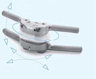
The TOPS System is a mechanical device that can be implanted into the spine at the affected segment to restore a controlled range of motion, facilitating extension, flexion, axial rotation, and lateral bending. The FDA has approved TOPS for people between 35 and 80 years old with degenerative spondylolisthesis (at one level between L3 to L5) and lumbar spinal stenosis.
Dr. Meyer, a neurosurgeon at the Atlantic Neuroscience Institute, was selected to participate in an FDA study of the TOPS System as a treatment for degenerative grade 1 spondylolisthesis and spinal stenosis. Dr. Meyer notes that the study presents patients with an opportunity to reap the benefits of this innovative device, including a restored range of motion, while correcting their spinal condition.
Spondylolisthesis Recovery Forecast and Long-Term Outlook
Recovery forecasts for grade 1 spondylolisthesis typically span three to six months. If surgery is required (which is rare), it may take six to 12 months to fully recover.
Non-Surgical Treatment Recovery
Cases of grade 1 spondylolisthesis that are successfully treated without surgery typically heal within three to six months, as mentioned above. Young patients may heal faster, while older patients may need more time to recover.
A spine specialist can evaluate your case of spondylolisthesis and provide a personalized recovery forecast based on your:
- Age
- Medical history
- Treatment plan
- Degree of slippage
Surgical Recovery Forecasts (Fusion vs. TOPS)
Most patients who undergo TOPS surgery for grade 1 spondylolisthesis return to their regular activities within around six weeks, while spinal fusion patients may need a year to recover. Fusion patients typically need to follow significant movement restrictions for around six to eight months following the procedure.
Long-Term Management and Preventing Progression
To manage grade 1 spondylolisthesis in the long-term, implement these lifestyle habits into your routine:
- Weight management
- Low-impact exercise, including core strengthening
- Stretching, focusing on the hamstrings and hips
- Annual check-ups with your spine specialist to facilitate early intervention
- Abstaining from smoking, which is detrimental to overall spine health
- Hydration and proper nutrition
FAQs
- Is grade 1 spondylolisthesis serious?
- Not typically, as it often has few to no symptoms
- How do you fix grade 1 spondylolisthesis?
- Physical therapy, activity adjustments, NSAIDs, and, in some cases, back bracing
- Can spondylolisthesis heal on its own?
- It can greatly improve with treatment, although the slippage can’t be reversed without surgery.
- How long does it take to recover from grade 1 spondylolisthesis?
- Three to six months, typically
- What triggers spondylolisthesis?
- Spinal degeneration, wear and tear, stress fractures, and genetic abnormalities are the most common triggers.